feature
The Practicality of the Non-Mydriatic Fundus Camera
This
relatively inexpensive diagnostic tool can greatly increase your imaging capabilities.
BY SHALOM KELMAN, M.D.
The non-mydriatic fundus camera has made great strides in the last couple of years, and it has developed into a diagnostic instrument many ophthalmology practices might find useful. At a fraction of the cost of some sophisticated camera systems, these cameras provide colorful, vivid images. I will share my experiences with this fundus camera, and the benefits and limitations associated with it.
|
|
|
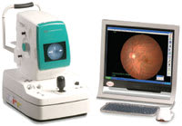
The Kowa fundus camera system's fail-safe "fire and forget" software prevents images from getting misplaced. |
Going Solo
When I was practicing in a university setting, I was accustomed to working with skilled, fundus photographers who readily could take photos during the course of an exam. When I decided to go into a private neuroophthalmology practice, I knew I needed a fundus camera that I could rapidly operate and did not interfere with patient flow. Given my limited manpower and financial constraints, I needed a more streamlined and economical solution. After doing some research, I purchased a non-mydriatic fundus camera (the Kowa Non-Myd a-D Fundus).
Based on my university experience, I initially assumed that my technician would take the photographs, and I would review afterwards. I soon discovered with the camera's ease-of-use, I could efficiently take photographs without adding significant time to the patient encounter. The camera's role had changed from an ancillary study to an extension of my slit-lamp and fundus examination.
Benefits
Setting up a fundus camera in your examination room requires only a modest investment in time and money. A digital fundus camera can create the same level of high-quality images as some more expensive, sophisticated diagnostic systems that you find in large practices or university settings. The color rendition of these cameras is outstanding, and details are extremely sharp in its images.
|
|
|
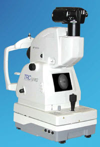
The TopCon TRC-NW6S features eight peripheral fixation points, as well as a built-in standard central fixation point, which provides fixed, reproducible fields for use in various study protocols. |
In my practice, I show patients images of their eyes during the exam to help explain pathology and evaluate progress of their diseases. I typically see patients with papilledema, optic neuritis and other diseases affecting the optic nerve. Rather than speak in general and abstract terms, I can point to the optic nerve photos and precisely detail the pathologic changes, and compare previous photographs to the current images. It creates visual evidence for the patient to view their own microaneurysms, hemorrhages and exudates associated with their diabetes than those shown in a schematic diagram or in other photographs.
After viewing their photos, patients feel like they can make more informed decisions regarding treatment and management. It is my impression that using the camera in this way has reduced misunderstandings and has enhanced relationships with my patients.
The camera's software allows for viewing of the stereo images without 3-D glasses or other visual aids. Taking disc stereo photographs is straightforward and does not require any advance training. This is a particularly powerful feature, which allows patients to observe optic disc changes that occur over time. For example, patients who have undergone successful optic nerve decompression surgery are thrilled to view improvements in their papilledema.
Before the advent of these cameras, stereo photos were difficult to obtain and almost impossible to show patients. A patient and physician can now simultaneously view the images and appreciate their significance.
Media opacities such as corneal scars or cataracts interfere with the clear fundus photo viewing. The resulting blurred photographs provide a powerful demonstration to the patient of the effect the cataract is having on their vision.
Organizing picture files is straightforward and mostly automated. The only data field the software requires is the patient's name. The date is automatically updated and all the patient files are placed in one location. Searching for previous photographs requires only typing in the first few letters of the patient's name.
Limitations
|
|
|
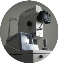
The Canon CR-DGi captures digital images that can be used with many different applications, such as telemedicine and electronic filing. |
The camera is not designed to take fluorescein fundus photographs. Although taking peripheral images is possible, the camera is optimized for photos of the posterior pole. The standard sizes are a wide field 45° image and a magnified 20° field. The internal fixation is sometimes difficult for patients to follow. For patients with poor vision, and who cannot see the fixation device, an external fixation device is available. However, I found it to be of limited use.
Almost all of my patients are dilated prior to photography, and I generally evaluate the fundus with a 78-diopter lens at the slit-lamp before taking photographs. On the few occasions that I have not dilated the pupil, image quality has suffered and is not up to par with the mydriatic images. Nevertheless, the images are of good quality and usable.
Coding and Photo Transfer
Coding for reimbursement of the images is straightforward. Medicare and other insurers accept a wide range of diagnoses for the 92060 fundus photography code, which can be found on the Medicare Web site. Reimbursement for the photographs is at a reasonably good level and allowed me to recoup my investment within a relatively short period of time.
The native images are encoded in a proprietary program that allows for various types of manipulation, but for transferring to other programs the JPEG encoded images are used. The JPEG images transfer to other software without loss of image quality. The digital images can be sent to third party insurers or colleagues via e-mail.
I routinely transfer images to PowerPoint for use in lectures and frequently e-mail images to referring physicians for their medical records. A color printer is provided as part of the package and produces high-quality images on glossy paper. However, I find that most physicians and patients prefer the digital images.
The Big Picture
In my practice, the non-mydriatic camera is mainly used for optic nerve imagery, and in particular, stereo images. The image quality allows for detailed images of the macula and posterior pole vessels. Any structure can be magnified to any level desired with the software.
Utilizing this camera has allowed me to evaluate pathologies more efficiently and to explain my patients' diseases more effectively.
Shalom Kelman, M.D., is the principal of Maryland Neuroophthalmology, LLC, located in Baltimore, Md. He can be e-mailed at skelman@comcast.net.
For Additional Information on Cameras
The following is a sampling of manufacturers that offer non-mydriatic fundus cameras in the North American market:
A Canon USA, Inc.
(800) 828-4040
www.usa.canon.com/eye-care
A Kowa-Optimed, Inc.
800-966-5692
www.kowa-usa.com
A TopCon America Corp.
(800) 223-1100
www.topcon.com


