Dry eye and ocular surface disease (OSD) are conditions I encounter frequently in my practice. Despite its prevalence,1 dry eye disease (DED) is multifactorial and highly variable, requiring a nuanced approach to address its diverse etiologies and presentations. Navigating the complexities of DED can be challenging, particularly when determining the optimal stepwise treatment plan for patients who have not responded to initial therapies.
However, regardless of its etiology, the inflammatory cycle that persists in DED involves tear film instability and hyperosmolarity, tissue damage, and some degree of corneal nerve dysfunction. The relationship between DED and the corneal nerves is complex, and there are clinical overlaps between DED and neurotrophic keratitis.2
Before we reach the stage where significant neurosensory abnormality is at play, we often have opportunities to intervene on key driving aspects of the DED presentation. Managing DED is not always straightforward, but identifying and treating its root causes is vital for maintaining the ocular health, vision, and quality of life of our patients.
Knowing the Background Causes of DED
The inflammatory processes behind chronic DED can be easily missed, and it is important to note that these processes often overlap and influence each other. For example, Demodex blepharitis or ocular rosacea can lead to meibomian gland dysfunction (MGD) and, consequently, evaporative DED.3 If we do not treat these underlying causes, other DED interventions are unlikely to succeed.
Indeed, chronic untreated or under-treated DED can have long-term consequences, including corneal nerve damage.4 Chronic DED can also contribute to issues like pterygia and Salzmann nodules,5,6 which can lead to further loss of corneal sensation, instigating a vicious cycle that reinforces itself and worsens both dry eye and corneal nerve damage.
Other conditions that may affect eyelid function or cause damage to periocular tissues can lead to neurotrophic keratitis. These include Bell’s palsy, ectropion, lagophthalmos following blepharoplasty, and damage following radiation to the head or neck, among others.
Given this complexity, it’s not uncommon to encounter patients who have been misdiagnosed or have received treatments that fail to address a key underlying issue. Identifying the root cause(s) of a multifactorial disease like DED can be challenging, but it is essential to focus on the specific drivers of the disease for each patient to deliver targeted and effective treatment. For example, while lifitegrast ophthalmic solution 5% (Xiidra; Bausch + Lomb) may be highly effective in treating surface-level inflammation, it cannot resolve eyelid margin disease on its own.
Putting Corneal Nerve Function in Context
Given the significant overlap of symptoms between DED and other OSD etiologies, a thorough patient history and detailed clinical examination are essential for an accurate diagnosis. Understanding the timing and duration of symptoms; identifying triggers such as prolonged computer use, dry environments, specific facial cleansers, or makeup brands; and assessing the effectiveness of prior treatments can provide valuable insights.7 This meticulous approach is especially crucial before cataract or refractive surgery, as untreated DED can exacerbate symptoms and lead to suboptimal visual outcomes postoperatively.8,9
Over time, I’ve found that the diagnostic process becomes something of an art. As we gain experience and begin to recognize patterns, we can more rapidly identify key contributing factors and masquerading conditions. For example, in this context, when I see a patient with marked signs of DED (diffuse punctate staining, low tear breakup time, etc.) but few symptoms, I think of neurotrophic keratitis. When I suspect this, I usually employ a wisp test—using a cotton swab with the fibers twisted into a point—to assess corneal sensitivity in the clinic.
Finding the Right Starting Point
If I see a patient who is obviously neurotrophic, I will skip through the first few steps I would normally employ and initiate them on cenegermin-bkbj (Oxervate; Dompé) immediately. However, in patients with DED whose presentations are more subtle, I will typically begin with a conservative treatment routine and see how they respond.
Conservative treatments often include ocular lubricants, eyelid hygiene and warm compresses, along with lifestyle or dietary modifications as needed. I make it a priority to ensure my patients are using high-quality products to minimize the risk of adverse reactions. For example, I recommend a foam cleanser like Eye Revive (Daily Practice), for a gentle yet effective approach to eyelid hygiene, and moist heat masks like Optase (Scope Eyecare) for warm compresses that retain consistent heat. Additionally, discussing skincare routines with patients is essential to help them identify safe options and avoid potentially harmful products. Many patients respond well to these conservative measures and may not require further intervention.
The Next Level
Further treatments are tailored to address the primary underlying causes of a patient’s DED. For MGD, in-office procedures such as thermal pulsation therapy or manual gland expression can help clear blocked glands and improve the health of the eyelid margin.10 Intense pulsed light therapy (Lumenis Optilight) has been shown to be a safe and effective treatment for MGD associated with ocular rosacea, leading to improved tear break-up time, reduced inflammation and significant relief from DED symptoms.11
To facilitate corneal healing, I make liberal use of cryopreserved amniotic membrane like Prokera or CAM360 (BioTissue), which promotes nerve regeneration and protects the ocular surface. Cryopreserved amniotic membranes have demonstrated efficacy in reducing the signs and symptoms of DED. Treatment duration is short and can have lasting benefits.12,13 I prefer the use of cryopreserved rather than dehydrated amniotic membranes because cryopreservation better preserves the matrix component that is responsible for much of the amniotic membrane’s therapeutic effects.14
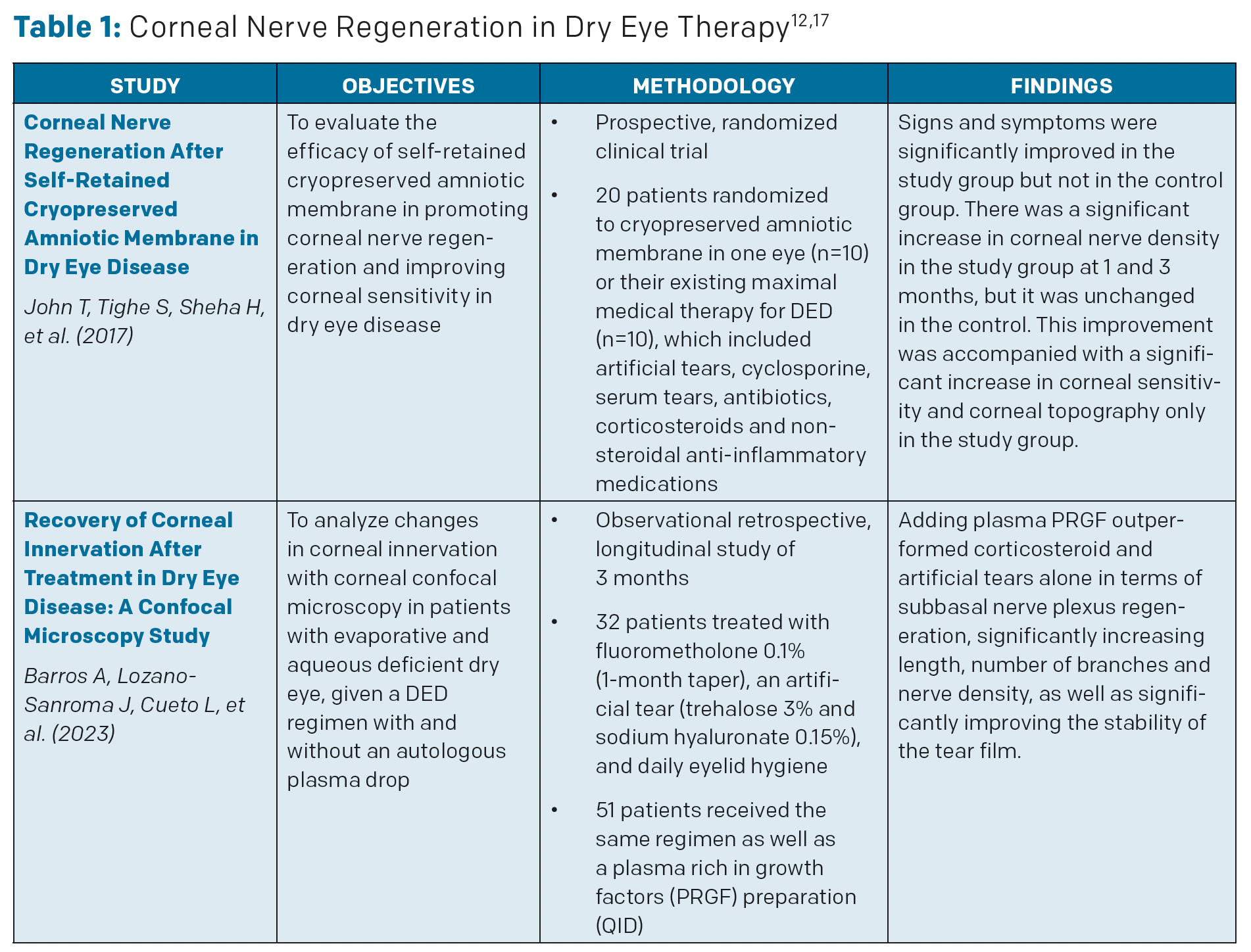
Autologous serum-based eye drops are another option that can help patients who have found insufficient relief from other therapies. Serum tears have demonstrated efficacy for corneal nerve regeneration in some patients,15 but a systematic review of randomized controlled trials found mixed efficacy results in DED.16 Because of this, and due to cost and logistical concerns, I seldom use autologous serum drops in my practice. Selected studies of corneal nerve regeneration in dry eye therapy can be found in Table 1.
Taking the Long View
A recent case underscores the complexity of managing dry eye and OSD. A gentleman visited our clinic after an astonishing 152 visits to his previous provider over the course of several years. He was self-referred to me as a refractory DED case, but upon examination, I identified multiple contributing factors, including epithelial basement membrane dystrophy, recurrent corneal erosions, and severe Demodex blepharitis.
Although he had undergone a superficial keratectomy, his relief was incomplete because these underlying issues had not been fully addressed. With the availability of lotilaner ophthalmic solution 0.25% (Xdemvy; Tarsus Pharmaceuticals), which specifically targets Demodex, we were able to successfully manage this significant contributor to his DED.
This case illustrates the multifaceted nature of diagnosing and treating DED and OSD. Effectively addressing the patient’s Demodex blepharitis allowed other treatments to take hold, restoring tear film homeostasis and promoting corneal healing. His treatment is currently ongoing.
Conclusions
Overall, it can help to think of treating DED as a marathon, not a sprint. If we pace ourselves and find the right points to intervene at the outset, we can often forestall the tissue damage, nerve dysfunction, and frustrations associated with longstanding DED.
It is also vital that we remain on the cutting edge of what we can offer to our patients by staying current with the literature, attending conferences, and connecting with our colleagues to learn what has been working for them and their patients. OM
References
1. Farrand KF, Fridman M, Stillman IÖ, Schaumberg DA. Prevalence of diagnosed dry eye disease in the United States among adults aged 18 years and older. Am J Ophthalmol. 2017;182:90-98. doi:10.1016/j.ajo.2017.06.033
2. Mead OG, Tighe S, Tseng SCG. Amniotic membrane transplantation for managing dry eye and neurotrophic keratitis. Taiwan J Ophthalmol. 2020;10(1):13-21. Published March 4, 2020. doi:10.4103/tjo.tjo_5_20
3. Narang P, Donthineni PR, D’Souza S, Basu S. Evaporative dry eye disease due to meibomian gland dysfunction: Preferred practice pattern guidelines for diagnosis and treatment. Indian J Ophthalmol. 2023;71(4):1348-1356. doi:10.4103/IJO.IJO_2841_22
4. Bron AJ, de Paiva CS, Chauhan SK, et al. TFOS DEWS II pathophysiology report [published correction appears in Ocul Surf. 2019 Oct;17(4):842]. Ocul Surf. 2017;15(3):438-510.
5. Linaburg T, Choi D, Bunya VY, Massaro-Giordano M, Briceño CA. Systematic review: Effects of pterygium and pingueculum on the ocular surface and efficacy of surgical excision. Cornea. 2021;40(2):258-267. doi:10.1097/ICO.0000000000002575
6. Roszkowska AM, Azzaro C, Calderone A, et al. Salzmann nodular degeneration in ocular and systemic diseases. J Clin Med. 2024;13(16):4900. Published August 20, 2024. doi:10.3390/jcm13164900
7. Amescua G, Ahmad S, Cheung AY, et al. Dry eye syndrome preferred practice pattern. Ophthalmology. 2024;131(4):P1-P49. doi:10.1016/j.ophtha.2023.12.041
8. Donthineni PR, Deshmukh R, Ramamurthy C, Sangwan VS, Mehta JS, Basu S. Management of cataract in dry eye disease: Preferred practice pattern guidelines. Indian J Ophthalmol. 2023;71(4):1364-1372. doi:10.4103/IJO.IJO_2807_22
9. Starr CE, Gupta PK, Farid M, et al. An algorithm for the preoperative diagnosis and treatment of ocular surface disorders. J Cataract Refract Surg. 2019;45(5):669-684. doi:10.1016/j.jcrs.2019.03.023
10. Tao JP, Shen JF, Aakalu VK, et al. Thermal pulsation in the management of meibomian gland dysfunction and dry eye: A report by the American Academy of Ophthalmology. Ophthalmology. 2023;130(12):1336-1341. doi:10.1016/j.ophtha.2023.07.009
11. Tashbayev B, Yazdani M, Arita R, Fineide F, Utheim TP. Intense pulsed light treatment in meibomian gland dysfunction: A concise review. Ocul Surf. 2020;18(4):583-594.
12. John T, Tighe S, Sheha H, et al. Corneal nerve regeneration after self-retained cryopreserved amniotic membrane in dry eye disease. J Ophthalmol. 2017;2017:6404918. doi:10.1155/2017/6404918
13. McDonald M, Janik SB, Bowden FW, et al. Association of treatment duration and clinical outcomes in dry eye treatment with sutureless cryopreserved amniotic membrane. Clin Ophthalmol. 2023;17:2697-2703. Published September 12, 2023. doi:10.2147/OPTH.S423040
14. Tighe S, Mead OG, Lee A, Tseng SCG. Basic science review of birth tissue uses in ophthalmology. Taiwan J Ophthalmol. 2020;10(1):3-12. Published March 4, 2020. doi:10.4103/tjo.tjo_4_20
15. Aggarwal S, Kheirkhah A, Cavalcanti BM, et al. Autologous serum tears for treatment of photoallodynia in patients with corneal neuropathy: Efficacy and evaluation with in vivo confocal microscopy. Ocul Surf. 2015;13(3):250-262. doi:10.1016/j.jtos.2015.01.005
16. Quan NG, Leslie L, Li T. Autologous serum eye drops for dry eye: Systematic review. Optom Vis Sci. 2023;100(8):564-571. doi:10.1097/OPX.0000000000002042
17. Barros A, Lozano-Sanroma J, Queiruga-Piñeiro J, et al. Recovery of corneal innervation after treatment in dry eye disease: A confocal microscopy study. J Clin Med. 2023;12(5):1841. Published February 25, 2023. doi:10.3390/jcm12051841


