


Preoperative planning for cataract surgery can be a time-consuming endeavor—particularly for the patient with astigmatism. But the ARGOS® Biometer with Image Guidance (Alcon) helps streamline the preoperative process while connecting the clinic data to the operating suite. It also features astigmatic management tools, including the LRI nomogram, to simplify astigmatism management.1 Scott LaBorwit, MD, and Kourtney Houser, MD, say that these capabilities—along with others—have made the ARGOS® a welcomed technology in their clinics.
What initially drew you to ARGOS®?
Scott LaBorwit, MD: My practice is dedicated solely to cataract surgeries, so it’s vital to have the most up-to-date diagnostic equipment. I initially looked at ARGOS® for the swept-source OCT, but quickly realized its speed of acquisition could benefit my clinic.2
With the additional ability to take measurements through dense cataracts,3 I can save time by not needing a contact A-scan. Studies showed statistically significant axial length measurement accuracy with ARGOS® for short and long eyes due to the sum of segments that use individual refractive indices to drive better predicable outcomes.4-5
Kourtney Houser, MD: I was first drawn to the easy image capture and biometry acquisition.1,2,3,6,7 Patients coming into the clinic for a cataract evaluation typically undergo evaluation for an hour, including workup and testing, so any technology that could shorten that time was beneficial. I have several patients with mature cataracts who would require an immersion A-scan for axial length acquisition with other devices that we can now bypass with ARGOS®.
How did ARGOS® change and improve your pre-op planning process?
Dr. LaBorwit: ARGOS’® ability to connect my clinic data to the operating suite was a gamechanger. It measures the [sitting] patient’s astigmatism and features of the iris and sclera, which are correlated by a digital marker. This allows a surgeon to confidently manage astigmatism without needing to account for torsional rotation when the patient is lying down, or twisting the microscope off the 180-degree axis.1
Dr. Houser: My preoperative planning process historically involved using one or two biometry devices to compare the consistency of measurements and to analyze the magnitude and direction of astigmatism. I would then manually input the data into various online calculators or nomograms to determine the best course of action for my patient’s vision. While I still verify magnitude, axis, and regularity of astigmatism with tomography, with ARGOS® I can now bypass the utilization of multiple biometers, online calculators, and nomograms during my workflow.1
How has ARGOS® positively impacted your surgical workflow and the workload of your technicians?
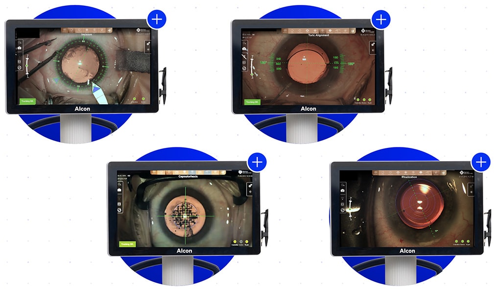
Dr. LaBorwit: The ARGOS® graphic interface and acquisition speed1-3 has enhanced my cataract clinic workflow, while decreasing the need for contact A-scans in many dense lenses. That data is then integrated into the EHR medical record, allowing me to evaluate and review it during consults or to create surgical plans in under a minute.1-3 It’s also imported to the LenSx® and ORA® to minimize work for my staff and to lessen transcription errors.1,8 It’s crucial during astigmatism management in the OR with a digital overlay on the Ngenuity® 3D heads-up display.
Dr. Houser: Technicians love the speed and ease of image capture and the significant reduction in need for immersion A-scans for axial length determination. The integrated planning software helps save time when constructing surgical plans, especially when considering astigmatism correction, and everything can be viewed on our network.1-3 I can now review a patient’s biometry before entering the exam lane and have a lens recommendation already formulated and graphically depicted if the patient wants to pursue astigmatism or presbyopia correction. In the operating room, I can feel confident with image-guided surgery, as ARGOS® provides an additional level of precision to our traditional manual markings.1
How has ARGOS® with Image Guidance impacted your clinic and practice as a whole?
Dr. LaBorwit: The digital marker is a gamechanger. The surgeon is engaged during surgery by using the pre-op clinical images of the patient, which are then overlapped with images obtained during femtosecond laser surgery (LenSx®) and the digital marker placement for the toric IOL rotation.9,10 This gives the surgeon certainty for each component of the toric management during cataract surgery.Overall, there is less stress for everyone—the surgeon, staff, and patients. My staff no longer needs to take time to input data or be worried about a transcription error impacting a patient’s outcomes permanently.1,8 Providers used to have to manually find and manage astigmatism, but their efforts were limited. ARGOS® has significantly elevated this surgeon’s confidence to deliver astigmatism correction of an exact axis.
Dr. Houser: The most impactful benefit of ARGOS® is its capacity to serve as a full surgical planning system instead of just a testing device in clinic.1,8 Its true value lies in its capacity to generate the best recommended option for astigmatism correction, whether it be a toric intraocular lens or a peripheral corneal relaxing incision, and also in its capability to transmit image registration information to the operating room.1
Image-guided cataract surgery is starting to raise the bar for refractive cataract surgery, which is what my patients are starting to expect. Being able to orient a lens or limbal relaxing incision on the precise axis of astigmatism increases my confidence that my patients will have minimal refractive error postoperatively.1 This also builds my confidence for offering and utilizing more presbyopia-correcting lenses, which are much more sensitive to residual astigmatism and refractive error.
Scott LaBorwit, MD, is an assistant professor at the Wilmer Eye Institute at The Johns Hopkins Hospital, as well as the founder of Select Eye Care with two locations in Maryland. He specializes in cataract and refractive surgery.
Kourtney Houser, MD, is an assistant professor in the department of ophthalmology at Duke University School of Medicine in Durham, N.C. She specializes in cataract surgery, corneal transplants, cornea and external disease, IOL complications, and refractive surgery.
References
- ARGOS® Biometer User Manual. 2019.
- Hussaindeen JR, Mariam EG, Arunachalam S, et al. Comparison of axial length using a new swept-source optical coherence tomography-based biometer. PLoS ONE. 2018;13(12):e0209356.
- Shammas HJ, Ortiz S, Shammas MC, et al. Biometry measurements using a new large-coherence-length swept-source optical coherence tomographer. J Cataract Refract Surg. 2016;42:50-61.
- Whang W, Yoo Y, Kang M, et al. Predictive accuracy of partial coherence interferometry and swept-source optical coherence tomography for intraocular lens power calculation. Sci Rep. 2018;8(1):13732.
- Wang L, Cao D, Weikert MP, Koch DD. Calculation of axial length using a single group refractive index versus using different refractive indices for each ocular segment. Theoretical Study and Refractive Outcomes. Ophthalmology. 2019:126(5):663-670.
- Tamaoki A, Kojima T, Hasegawa A, et al. Clinical evaluation of a new swept-source optical coherence biometer that uses individual refractive indices to measure axial length in cataract patients. Ophthalmic Res. 2019;19:1-13.
- ZEISS IOLMaster 700 510k Submission. 2015.
- VERION® Reference Unit User Manual Part II. 2017.
- VERION® Digital Marker L User Manual. 2017.
- VERION® Digital Marker M User Manual. 2020.
Important Safety Information
The ARGOS® Biometer with Image Guidance by Alcon® Caution: Federal (USA) law restricts this device to the sale by or on the order of a physician.
Indications: ARGOS® is a non-invasive, non-contact biometer based on swept-source optical coherence tomography (SS-OCT). The device is intended to acquire ocular measurements as well as perform calculations to determine the appropriate intraocular lens (IOL) power and type for implantation during intraocular lens placement.
Intended Use: The Reference Image functionality is intended for use as a preoperative and postoperative image capture tool. It is intended for use by ophthalmologists, physicians, and other eye-care professionals and may only be used under the supervision of a physician.
Warnings and Precautions:
• Only properly trained personnel with experience may operate the device and control software and interpret the results.
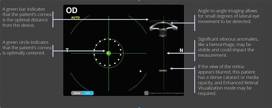
• Factors that influence the measurement of patient’s eyes are listed in the User Manual (Table 1): pseudophakic eye, wearing contact lenses, fixation problem, cornea opacity, non-intact cornea, refractive surgery, blood in the vitreous humor, retinal detachment, keratoconus, asteroid hyalosis, ambient light in the room, and deformation of the corneal shape. Please consider the guidance provided in Table 1 when you encounter these factors.
• Optical Radiation - This device is equipped with a Class 1 laser light source.
ATTENTION: Refer to the ARGOS® User Manual for a complete description of proper use and maintenance, optical and technical specifications, as well as a complete list of warnings and precautions.
© 2024 Alcon Inc. 05/24 US-ARB-2400018.
This content is supported by
